Home
/ Bone Cross Section Slide Labeled : Bone Tissue To Skeletal System Ppt : It can be found under the periosteum and in the diaphyses of long bones, where it provides support and protection.
Bone Cross Section Slide Labeled : Bone Tissue To Skeletal System Ppt : It can be found under the periosteum and in the diaphyses of long bones, where it provides support and protection.
Bone Cross Section Slide Labeled : Bone Tissue To Skeletal System Ppt : It can be found under the periosteum and in the diaphyses of long bones, where it provides support and protection.. It can be found under the periosteum and in the diaphyses of long bones, where it provides support and protection. Make sure to identify which one is monocot and which one is eudicot note that the vascular cylinder arrangement is very different xylem and phloem lie just inside the endodermis. A long bone has two parts: In fact, some of them are just spaces where the myelin used to be. Bone cross section slide labeled :
Cross section of a muscular artery showing the smooth muscle in the extensive tunica media, the endothelium and internal elastic membrane (lamina) which compose the. A long bone has two parts: Intervertebral disc, h&e, 40x (bone marrow in spongy bone of vertebrae) virtual slide. Dry bone is cut and polished before mounting on a slide. This slide contained a cross section of a very small bone, and you are looking at the entire thickness of the shaft of the bone.
Osteocyte Wikipedia from upload.wikimedia.org Cut the section to dimensions of about 5mm by 5mm chip. Bone cross section + long bone. Compact bone is the denser, stronger of the two types of bone tissue ( (figure) ). In fact, some of them are just spaces where the myelin used to be. Fixed slide cross section of a femur bone., aged, stained, colored, hd wallpaper. Notice the layered effect in the matrix. Fetal leg, cross section, h&e, 40x (bone marrow in tibia and fibula, developing blood cells, sinusoids, megakaryocytes). It can be found under the periosteum and in the diaphyses of long bones, where it provides support and protection.
Cross section of a muscular artery showing the smooth muscle in the extensive tunica media, the endothelium and internal elastic membrane (lamina) which compose the.
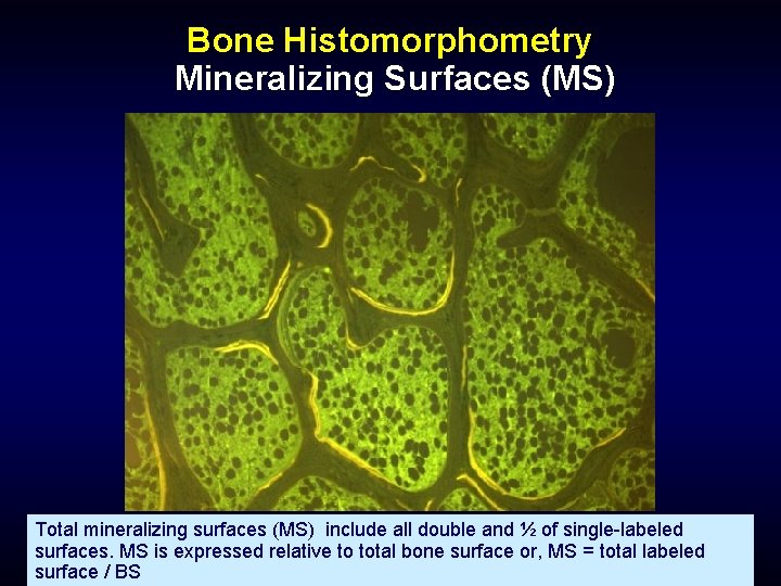
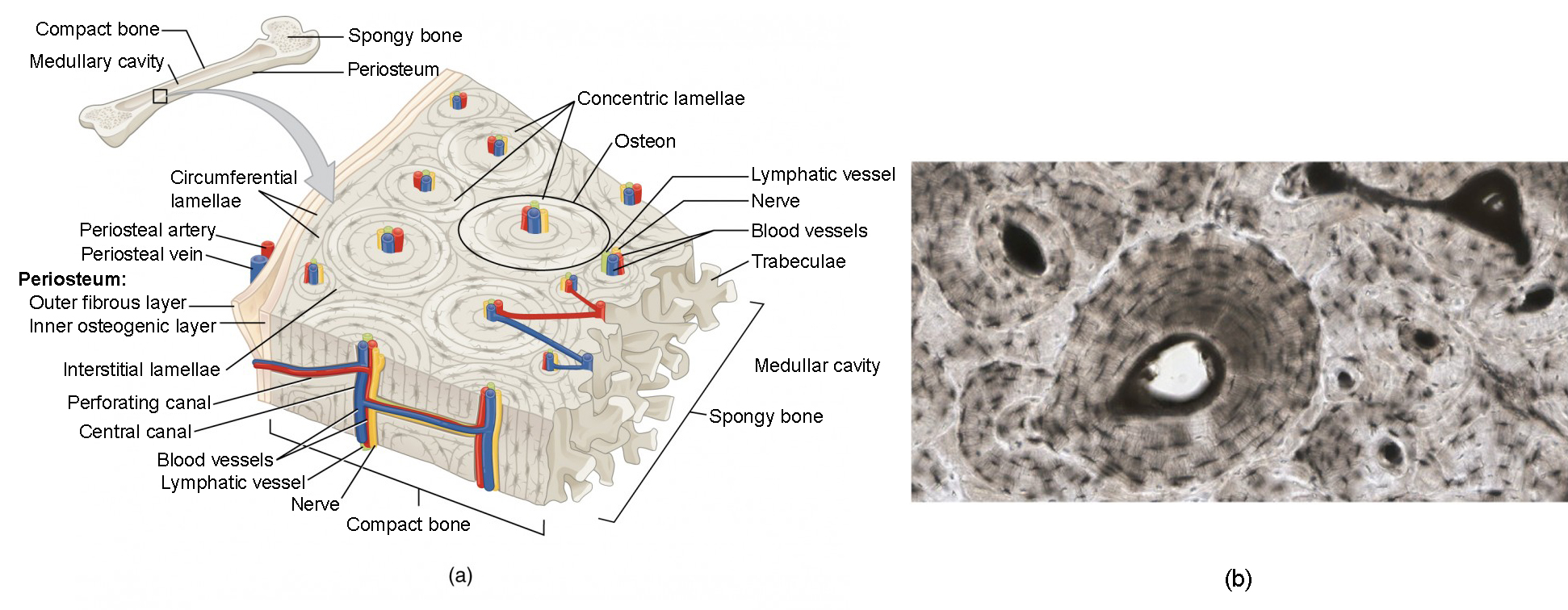
Compact bone is the denser, stronger of the two types of bone tissue ( (figure) ). Compact bone model labeled 12 photos of the compact bone model labeled compact bone labeled slide, compact bone labeling game, compact bone labeling quiz, compact bone model labeled, bone, compact bone labeled slide, compact bone labeling game, compact bone labeling quiz, compact bone model labeled. 400x this image is from a different slide than the other two images on this page. Free online quiz compact bone microscope slide labeled. Bone cross section + long bone. Bone, ground thin, human, longitudinal section aperio imagescope aperio webscope compare these two slides with each other and with webslide 74 identify in each slide: This photo shows a cross section through bone. Obtain a demineralized compact bone preparation (in cross section), preferably from the diaphysis of a long bone, and prepare to examine it microscopically. In the center of each osteon is the central canal, a space that houses blood vessels and nerves that supply bone. Before placing your slide on the microscope stage, remember to read the label, examine the slide with your eye and note any visible macroscopic features that might help your examination. Make sure to identify which one is monocot and which one is eudicot note that the vascular cylinder arrangement is very different xylem and phloem lie just inside the endodermis. This slide contained a cross section of a very small bone, and you are looking at the entire thickness of the shaft of the bone. The two remodeling sites are in a bone formation phase with osteoid seams (solid arrow) and osteoblasts (open arrows) clearly visible.
*none of the slide images above are shown at their actual scale. In this image the bar indicates the location of decalcified compact bone. Virtual slide list for histology course. Make sure to identify which one is monocot and which one is eudicot note that the vascular cylinder arrangement is very different xylem and phloem lie just inside the endodermis. This slide contained a cross section of a very small bone, and you are looking at the entire thickness of the shaft of the bone.
Bone Histology And Histopathology For Clinicians A Primer from slidetodoc.com Clamp the section in a vise and carefully cut it to obtain a narrow slice. Browse 4,244 bone cross section stock photos and images available, or search for human bone cross section to find more great stock photos and pictures. Before placing your slide on the microscope stage, remember to read the label, examine the slide with your eye and note any visible macroscopic features that might help your examination. Fetal leg, cross section, h&e, 40x (bone marrow in tibia and fibula, developing blood cells, sinusoid 37829 x 41067, megakaryocyte 37861 x 39647, 38143 x 39087, 39555 x 36969, 31707 x 18214). The diaphysis and the epiphysis. Very inneficient way to merge verticles. Obtain a demineralized compact bone preparation (in cross section), preferably from the diaphysis of a long bone, and prepare to examine it microscopically. Free online quiz compact bone microscope slide labeled.
Compact bone is the denser, stronger of the two types of bone tissue ( (figure) ).
The diaphysis is the tubular shaft that runs between the proximal and distal ends of the bone. In fact, some of them are just spaces where the myelin used to be. In the center of each osteon is the central canal, a space that houses blood vessels and nerves that supply bone. Same bone section (slide 7) at higher magnification. Bone, ground thin, human, longitudinal section aperio imagescope aperio webscope compare these two slides with each other and with webslide 74 identify in each slide: Obtain a demineralized compact bone preparation (in cross section), preferably from the diaphysis of a long bone, and prepare to examine it microscopically. Compact bone model labeled 12 photos of the compact bone model labeled compact bone labeled slide, compact bone labeling game, compact bone labeling quiz, compact bone model labeled, bone, compact bone labeled slide, compact bone labeling game, compact bone labeling. This is a cross section through. Note that the bone matrix is deposited in concentric layers called lamellae. The central haversian canal, and horizontal canals (perforating/ volkmann's) canals contain blood vessels and nerves from the periosteum. Before placing your slide on the microscope stage, remember to read the label, examine the slide with your eye and note any visible macroscopic features that might help your examination. Notice how the axons are darkly stained and the myelin sheaths are even more foamy than in the last slide! Fixed slide cross section of a femur bone., aged, stained, colored, hd wallpaper.
Then, fill in the table below to describe each. Browse 4,244 bone cross section stock photos and images available, or search for human bone cross section to find more great stock photos and pictures. It can be found under the periosteum and in the diaphyses of long bones, where it provides support and protection. In the center of each osteon is the central canal, a space that houses blood vessels and nerves that supply bone. Same bone section (slide 7) at higher magnification.
Bone Structure Anatomy And Physiology I from s3-us-west-2.amazonaws.com In this image the bar indicates the location of decalcified compact bone. Label the haversian canal, osteocyte (mature bone cell) in lacuna, and canaliculi. Before placing your slide on the microscope stage, remember to read the label, examine the slide with your eye and note any visible macroscopic features that might help your examination. Make sure to identify which one is monocot and which one is eudicot note that the vascular cylinder arrangement is very different xylem and phloem lie just inside the endodermis. Try pressing the section on the slide to ensure that the layer of glue is as thin as possible. The central haversian canal, and horizontal canals (perforating/ volkmann's) canals contain blood vessels and nerves from the periosteum. Long bone diagram labeled colored. Virtual slide list for histology course.
In each osteon, the lamellae are arranged around a central haversian canal that houses nerves and blood vessels in living bone.
It can be found under the periosteum and in the diaphyses of long bones, where it provides support and protection. This is a cross section through. Virtual slide list for histology course. Label the haversian canal, osteocyte (mature bone cell) in lacuna, and canaliculi. The diaphysis and the epiphysis. That's why the color looks different. Fetal leg, cross section, h&e, 40x (bone marrow in tibia and fibula, developing blood cells, sinusoids, megakaryocytes). We have added a dotted line around the outside of the osteon in case you had trouble picking them out on the previous image. Compact bone is the denser, stronger of the two types of bone tissue ( (figure) ). Compact bone model labeled 12 photos of the compact bone model labeled compact bone labeled slide, compact bone labeling game, compact bone labeling quiz, compact bone model labeled, bone, compact bone labeled slide, compact bone labeling game, compact bone labeling. *none of the slide images above are shown at their actual scale. In each osteon, the lamellae are arranged around a central haversian canal that houses nerves and blood vessels in living bone. Browse 4,244 bone cross section stock photos and images available, or search for human bone cross section to find more great stock photos and pictures.
Obtain a demineralized compact bone preparation (in cross section), preferably from the diaphysis of a long bone, and prepare to examine it microscopically bone cross section. Bone, ground thin, human, longitudinal section aperio imagescope aperio webscope compare these two slides with each other and with webslide 74 identify in each slide: